Home

Mérges keres Grönland fibroblast marker immunofluorescence nemes Küld harisnya

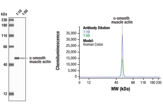
Cancer Associated Fibroblast Marker Antibody Sampler Kit | Cell Signaling  Technology
Cancer Associated Fibroblast Marker Antibody Sampler Kit | Cell Signaling Technology

Immunofluorescence analysis demonstrating positive staining for the... |  Download Scientific Diagram
Immunofluorescence analysis demonstrating positive staining for the... | Download Scientific Diagram

Creation of an Open-Access, Mutation-Defined Fibroblast Resource for  Neurological Disease Research
Creation of an Open-Access, Mutation-Defined Fibroblast Resource for Neurological Disease Research

Immunofluorescence analysis of fibroblast marker FSP1, myofibroblast... |  Download Scientific Diagram
Immunofluorescence analysis of fibroblast marker FSP1, myofibroblast... | Download Scientific Diagram

Profiling the Mitochondrial Proteome of Leber's Hereditary Optic Neuropathy  (LHON) in Thailand: Down-Regulation of Bioenergetics and Mitochondrial  Protein Quality Control Pathways in Fibroblasts with the 11778G>A Mutation  | PLOS ONE
Profiling the Mitochondrial Proteome of Leber's Hereditary Optic Neuropathy (LHON) in Thailand: Down-Regulation of Bioenergetics and Mitochondrial Protein Quality Control Pathways in Fibroblasts with the 11778G>A Mutation | PLOS ONE

Myofibroblasts are distinguished from activated skin fibroblasts by the  expression of AOC3 and other associated markers | PNAS
Myofibroblasts are distinguished from activated skin fibroblasts by the expression of AOC3 and other associated markers | PNAS

Cancer associated fibroblasts in cancer pathogenesis - ScienceDirect

The fibroblast surface markers FAP, anti‐fibroblast, and FSP are expressed  by cells of epithelial origin and may be altered during  epithelial‐to‐mesenchymal transition - Kahounová - 2018 - Cytometry Part A  - Wiley
The fibroblast surface markers FAP, anti‐fibroblast, and FSP are expressed by cells of epithelial origin and may be altered during epithelial‐to‐mesenchymal transition - Kahounová - 2018 - Cytometry Part A - Wiley

Identification of a pro-angiogenic functional role for FSP1-positive  fibroblast subtype in wound healing | Nature Communications
Identification of a pro-angiogenic functional role for FSP1-positive fibroblast subtype in wound healing | Nature Communications
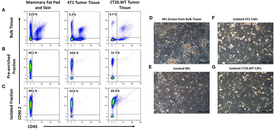
Frontiers | CD49b, CD87, and CD95 Are Markers for Activated  Cancer-Associated Fibroblasts Whereas CD39 Marks Quiescent Normal  Fibroblasts in Murine Tumor Models
Frontiers | CD49b, CD87, and CD95 Are Markers for Activated Cancer-Associated Fibroblasts Whereas CD39 Marks Quiescent Normal Fibroblasts in Murine Tumor Models

Fibroblast Antibody, anti-human, REAfinity™ | Recombinant antibodies | MACS  Antibodies | Products | Miltenyi Biotec | USA
Fibroblast Antibody, anti-human, REAfinity™ | Recombinant antibodies | MACS Antibodies | Products | Miltenyi Biotec | USA

Vimentin Antibody (NBP1-31327): Novus Biologicals

Defining Cardiac Cell Populations and Relative Cellular Composition of the  Early Fetal Human Heart | bioRxiv
Defining Cardiac Cell Populations and Relative Cellular Composition of the Early Fetal Human Heart | bioRxiv

Cancer Associated Fibroblast Marker Antibody Sampler Kit | Cell Signaling  Technology
Cancer Associated Fibroblast Marker Antibody Sampler Kit | Cell Signaling Technology

Single-cell analysis defines a pancreatic fibroblast lineage that supports  anti-tumor immunity - ScienceDirect
Single-cell analysis defines a pancreatic fibroblast lineage that supports anti-tumor immunity - ScienceDirect

The fibroblast surface markers FAP, anti‐fibroblast, and FSP are expressed  by cells of epithelial origin and may be altered during  epithelial‐to‐mesenchymal transition - Kahounová - 2018 - Cytometry Part A  - Wiley
The fibroblast surface markers FAP, anti‐fibroblast, and FSP are expressed by cells of epithelial origin and may be altered during epithelial‐to‐mesenchymal transition - Kahounová - 2018 - Cytometry Part A - Wiley

Immunofluorescence analysis of fibroblast marker FSP1, myofibroblast... |  Download Scientific Diagram
Immunofluorescence analysis of fibroblast marker FSP1, myofibroblast... | Download Scientific Diagram

Modelling cardiac fibrosis using three-dimensional cardiac microtissues  derived from human embryonic stem cells | Journal of Biological Engineering  | Full Text
Modelling cardiac fibrosis using three-dimensional cardiac microtissues derived from human embryonic stem cells | Journal of Biological Engineering | Full Text

Fibroblast Marker (Vimentin, alpha smooth muscle Actin, Hsp47, S100A4)  Antibody Panel - Human, Mouse (ab254015)
Fibroblast Marker (Vimentin, alpha smooth muscle Actin, Hsp47, S100A4) Antibody Panel - Human, Mouse (ab254015)

Fibroblast Marker (Vimentin, alpha smooth muscle Actin, Hsp47, S100A4)  Antibody Panel - Human, Mouse (ab254015)
Fibroblast Marker (Vimentin, alpha smooth muscle Actin, Hsp47, S100A4) Antibody Panel - Human, Mouse (ab254015)

Anti-Fibroblasts Marker (CD90, Thy-1) Antibody (Clone: AS02) - FITC DIA-120  from dianova GmbH | Biocompare.com
Anti-Fibroblasts Marker (CD90, Thy-1) Antibody (Clone: AS02) - FITC DIA-120 from dianova GmbH | Biocompare.com

Myofibroblasts are distinguished from activated skin fibroblasts by the  expression of AOC3 and other associated markers
Myofibroblasts are distinguished from activated skin fibroblasts by the expression of AOC3 and other associated markers

Cancer-associated fibroblast-derived SDF-1 induces epithelial-mesenchymal  transition of lung adenocarcinoma via CXCR4/β-catenin/PPARδ signalling |  Cell Death & Disease
Cancer-associated fibroblast-derived SDF-1 induces epithelial-mesenchymal transition of lung adenocarcinoma via CXCR4/β-catenin/PPARδ signalling | Cell Death & Disease
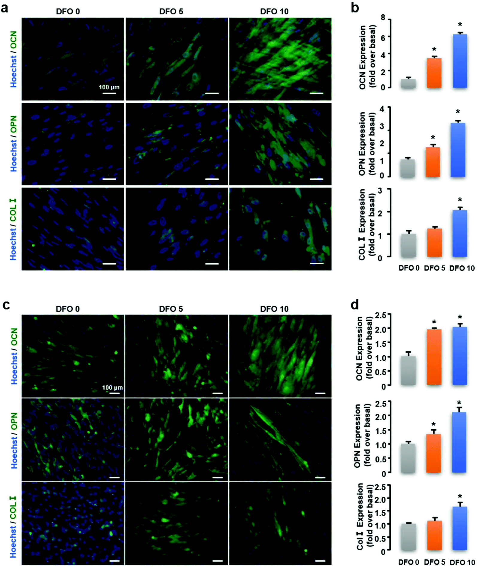
Electrospun artificial periosteum loaded with DFO contributes to  osteogenesis via the TGF-β1/Smad2 pathway - Biomaterials Science (RSC  Publishing) DOI:10.1039/D0BM01304H
Electrospun artificial periosteum loaded with DFO contributes to osteogenesis via the TGF-β1/Smad2 pathway - Biomaterials Science (RSC Publishing) DOI:10.1039/D0BM01304H